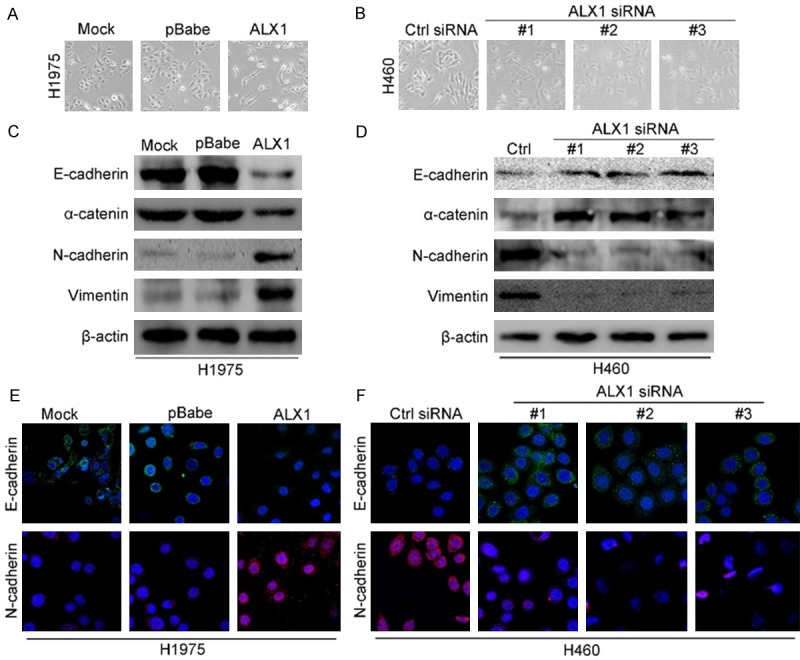
Figure 4.

ALX1 induces EMT in lung cancer cells. A. Micrographs showing the morphology of H1975 with overexpression of ALX1 and corresponding control cells. B. Micrographs showing the morphology of H460 with silencing ALX1 and corresponding control cells. C. Western blot analysis of the expression of the epithelial cell marker E-cadherin, alpha-catenin and the mesenchymal cell markers vimentin, N-cadherin in H1975 cell overexpressing ALX1. D. Western blot analysis of the expression of EMT markers in H1975 cell silencing ALX1. E. Immunofluorescence images of EMT markers in H1975 overexpressing ALX1. F. Immunofluorescence images of EMT markers in H460 silencing ALX1.
