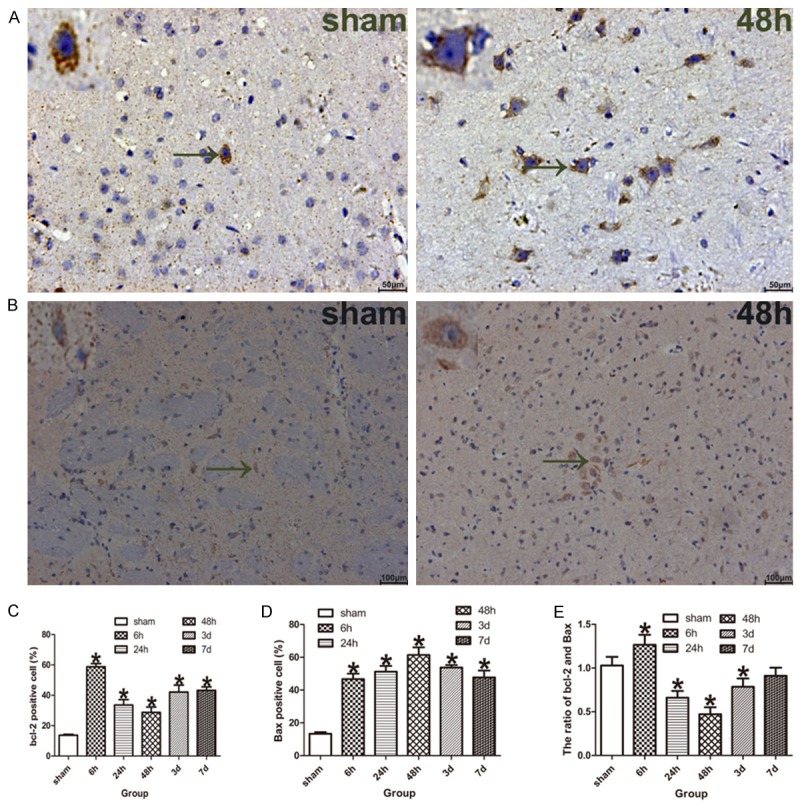
Figure 4.

Apoptosis-related proteins (bcl-2 and Bax) were measured by immunohistochemistry. A. The bcl-2 positive neurons in the ischemic penumbra of tMCAO model rats, the black arrow indicate bcl-2 positive neuron. B. The Bax positive neurons in the ischemic penumbra of tMCAO rats, the black arrow indicate Bax positive neuron. C. The bar graph shows the percent of bcl-2 positive neurons was increased in the model rats compared with the sham-operated rats (n = 5 for each group). D. The bar graph shows the percent of Bax positive neurons was increased in the model rats compared with the sham-operated rats (n = 5 for each group). E. The bar graph shows the ratio of bcl-2/Bax was increased at ischemia/reperfusion 6 h, while decreased at ischemia/reperfusion 24 h, 48 h, 3 and 7 d of the model rats compared with the sham-operated rats (n = 5 for each group). Data are expressed as mean ± SD. *P < 0.05 versus sham-operated group.
