
Figure 2.
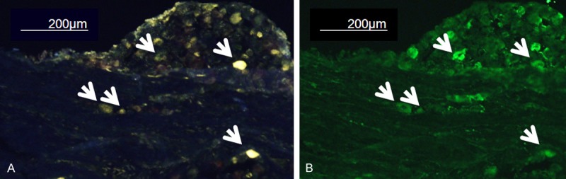
Representative immunofluorescence image of L-2 DRG. (A) FG-labeled image and (B) CGRP-immunostain imaging from the same slice. Arrows indicate the CGRP-immunoreactive FG-labeled DRG neurons. DRG: dorsal root ganglia, FG: Fluorogold (neurotracer), CGRP: calcitonin gene-related neuropeptide (biomarker for inflammatory pain).