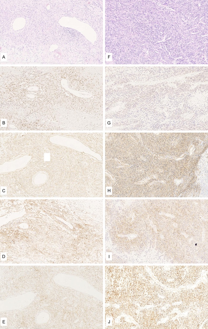
Figure 2.

H&E and immunohistochemical studies of SFTs/HPCs. A-E: Case #1, benign SFT. F-J: Case #43, malignant SFT. A: SFT component was composed of spindle or oval-shaped tumor cells intervening with different thickness collagen, some were broad bands of hyalinized collagen. Tumor cells arranged sparse and showed hemangiopericytoma-like growth pattern. The cellular pleomorphism was invisible; H&E, 100×; B: Strong staining and diffuse nuclear positive (4+) for STAT6, 100×; C: Positive for CD34, 100×; D: Positive for CD99, 100×; E: Positive for Bcl-2, 100×; F: SFT hypercellular tumor cells arranged in hemangiopericytoma-like growth pattern. Tumor lesions showed increased mitoses (10 mitoses per 10 HPF), variable cytological atypia and dark stained nuclei; H&E, 100×; G: Cytoplasm positive for STAT6, 100×; H: Positive for CD34, 100×; I: Positive for CD99, 100×; J: Positive for Bcl-2, 100×.
