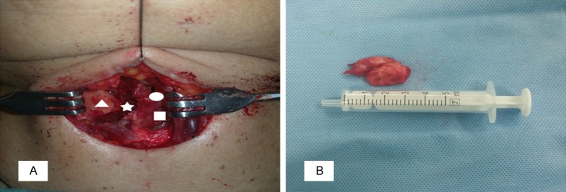
Figure 3.

Intraoperative findings. A. View of surgical cavity, it showed that the mass was excised completely from the right side of the paraglottic space (asterisk), the paries medialis of the thyroid cartilage (triangle), the right ventricular band (circular) and the right vocal cord (square) were exposed. B. A part of the resected mass was whitish-grey in color, measured 3.5 cm × 3.0 cm × 3.0 cm in total.
