
Figure 4.
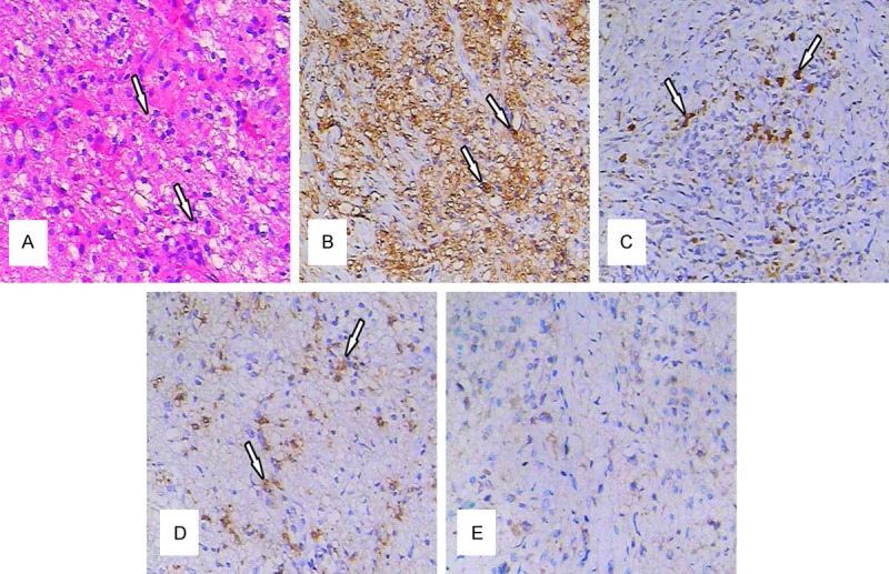
(A) Histopathological findings (H&E staining): The normal lymph node architecture was altered by a massive sinusoidal dilation of histiocytes in the inflammatory background (original magnification ×200). (B) Immunohistological findings: Some of the histiocytes exhibited S-100 (+) (original magnification ×200). (C) Immunohistological findings: Some of the histiocytes exhibited CD68 (+) (original magnification ×200). (D) Histiocytes exhibited immunoreactivity for lysozyme (+) (original magnification ×200) and (E) CD1a (-) (original magnification ×200).