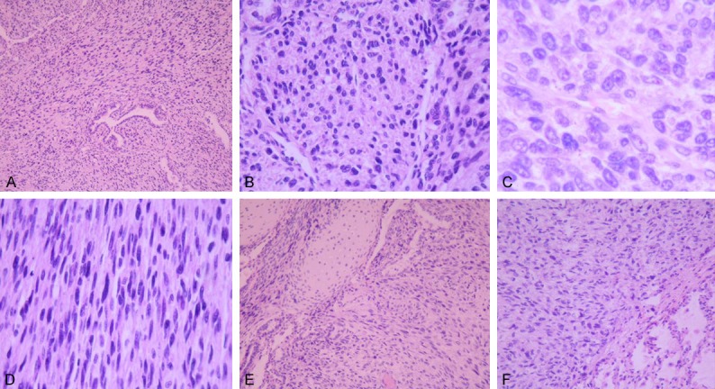
Figure 2.

A: The tumor mass demonstrated an aggregation of primitive cells with a round or orbicular-ovate shape and with residual mature bronchioles. B: (medium magnification of a) Cells were medium sized and had little cytoplasm, and the nuclei were round or orbicular-ovate and deeply stained. The cells were highly malignant. C: (high magnification of a) The cytoplasm contained vacuoles; the nuclei were deeply stained; and the nucleolus was clearly visible. Karyokinesis was active. D: A fibrosarcoma-like lesion was interwoven with spindle-shaped cells and short spindle-shaped cells. E: A malignant fibrous histiocytoma contained well-differentiated cartilage. F: A junction between the tumor and lung tissue. The right comprises malignant tumor cells, while the left comprises normal lung tissue.
