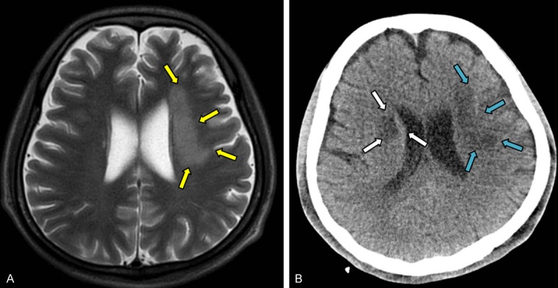
Figure 2.

(A) MRI of the brain showed a medium-size focal area of cerebral infarction near the left ventricle (yellow arrow). (B) A follow-up CT scan of the brain showed a newly emerged area of cerebral infarction near the right ventricle (white arrow) in addition to the prior cerebral infarction area (blue arrow) (B).
