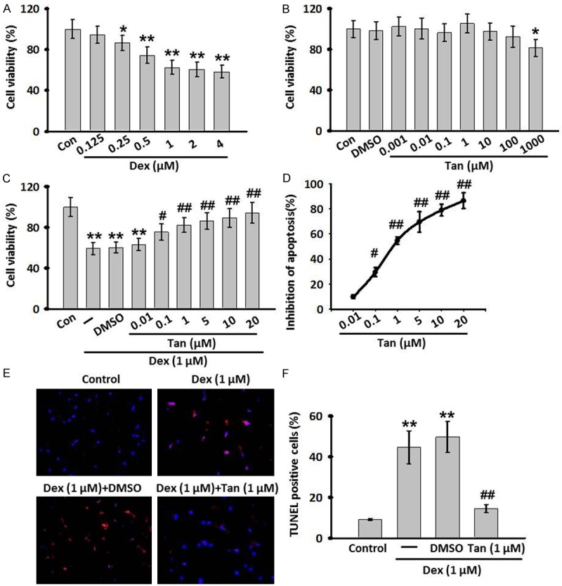
Figure 1.

Cell viability response to various concentrations of dexamethasone (Dex) treatment and the effects of Tan omDex-induced cell injury. (A, B) MC3T3-E1 cells were treated with various contractions of Dex (0.125-4 μM) (A) or Tanshinone IIA (Tan, 0.001-1000 μM) (B) for 24 h. Cell viability was determined by MTT assay. (C) The Dex induced decrease in MC3T3-E1 cells viability was improved by Tan at various concentrations. (D) The concentraction-response cruve of anti-apoptotic effect of Tan in MC3T3-E1 cells (IC50=9.646 μM). (E) TdT-mediated dUTP nick-end labeling (TUNEL) (red) and DAPI staining (blue) of MC3T3-E1 cells following co-incubation of Dex and Tan for 24 h. (F) The percentage of TUNEL positive cells was calculated. All data are presented as mean ± SEM. *P<0.05, **P<0.01 vs. control; #P<0.05, ##P<0.01 vs. Dex treatment alone, n=6.
