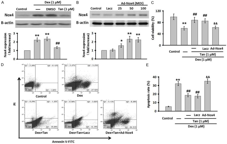
Figure 5.
Tan attenuated Dex-induced cell apoptosis through inhibiting Nox4 expression. A. MC3T3-E1 cells were co-incubated with Dex and Tan for 24 h. The expression of Nox4 was determined by western blot. B. The cells were infected with Ad-Nox4 for different MOI (25, 50, 100 MOI) or Laczfor 24 h. The expression of Nox4 was detected by western blot. C. Nox4 adenovirus (Ad-Nox4, 50 MOI) was transfected for 24 h in prior to the co-incubation of MC3T3-E1 cells with Dex and Tan. Cell viability was tested by MTT assay. D. Cell apoptosis was determined by Annexin V/PI staining. E. Quantitative analysis of the percentage of apoptotic cells. Data were presented asmean ± SEM. **P<0.01 vs. control; ##P<0.01 vs. Dex; &&P<0.01 vs. Tan + Dex, n=4.

