Figure 2.

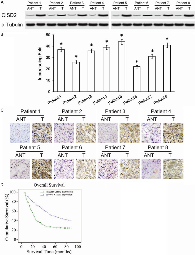
Overexpression of CISD2 mRNA and protein in primary HCC tissues. A. Western blotting of CISD2 protein expression in eight pairs of matched HCC cancer (T) and adjacent nontumor liver tissues (ANT). B. Average T/N ratios of CISD2 mRNA expression in paired liver cancer (T) and adjacent noncancerous tissues (N) were quantified using qPCR and normalized against α-Tubulin. Error bars represent the standard deviation of the mean (SD) calculated from three parallel experiments. C. Immunohistochemical assay of CISD2 protein expression in eight pairs of HCC tissues. D. Kaplan-Meier curves with univariate analyses (log-rank) for HCC patients with low CISD2 expression versus high CISD2 expression. The cumulative 5-year survival rate was 46.2% in the low CISD2 protein expression group (n=87), but only 24.5% in the high-expression group (n=109) *P<0.05.
