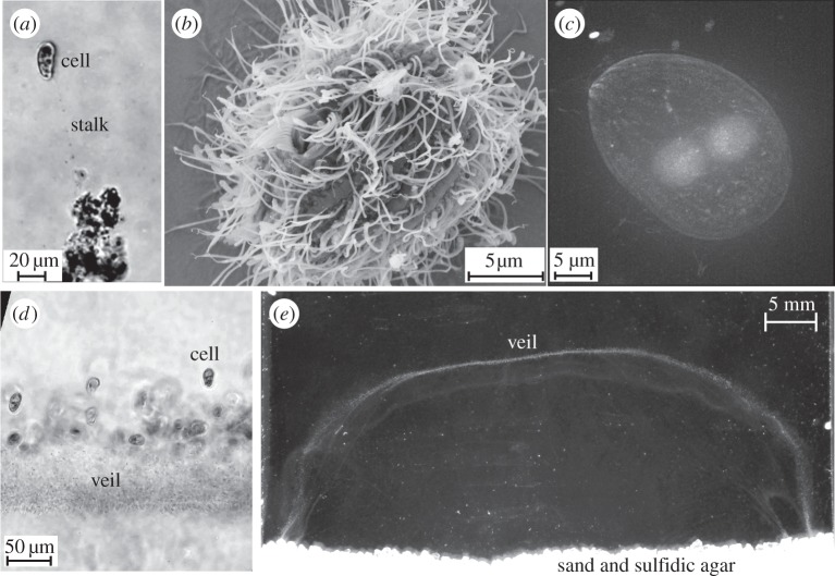
Figure 2.
Uronemella is a genus of ciliates. (a) Individual cells exude a thin mucous stalk. Several small particles can be seen stuck to this stalk. (b) An electron micrograph shows that individual cells are covered in several hundred cilia. The cell uses these cilia to pull water past the cell. Like the T. majus cell shown in figure 1b, the cell size has shrunk considerably as a result of desiccating the cells. (c) DAPI stained Uronemella overlain on dark field. Two spherical inclusions show the nuclei. (d) A front of Uronemella cells are shown with a veil. As the stalks of neighbouring cells become entwined, they form a veil. Note that nearly all of the cells are attached on the same side of the front. (e) An Uronemella veil grown between two microscope slides separated by 1 mm. The veil appears as a white U-shaped line. One millimolar sulfide diffusing from agar at the base of the chamber mixes with oxygen in the media, providing the sole energy source for the enrichment culture.

