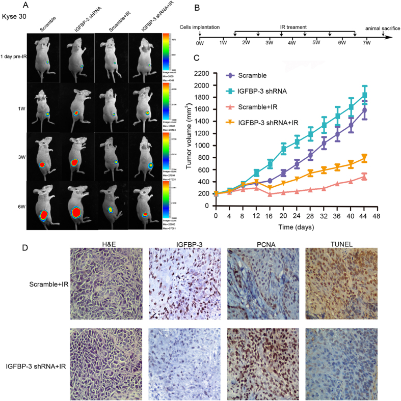
Figure 3. Silence of IGFBP-3 inhibits the therapeutic effect of IR on ESCC cell xenografts.
Kyse30-IGFBP-3shRNA and Kyse30-vector cells (3 × 10⁶) were injected s.c. into the lower limbs of athymic nude mice, respectively. When the volume of a transplanted tumor reached 200 mm3, mice were treated with a 6 Gy dose of IR. (A) Bioluminescent images of ESCC tumor xenografts was applied to detected the tumor size on 1day before IR treatment, 1 week, 3 weeks and 6 week after implantation of ESCC cells. (B) The procedure chart was shown. (C) The tumor volume of xenografts was measured with calipers every 3 days for a total of 35–44 days .The values represent mean tumor volume ± SE. (D) Representative images showing xenograft tumors in null mice from control and Kyse30-IGFBP-3 shRNA cells after IR treatment. H&E staining, IHC staining of IGFBP-3 and PCNA, and TUNEL assays were performed.

