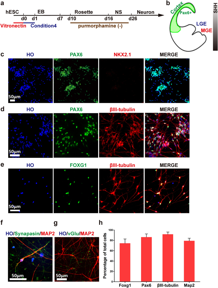
Figure 3. Directed differentiation of hPSCs to cortical neurons in a xeno-free system.
(a) Timeline of direct differentiation of hPSCs to cortical neurons in a xeno-free system by using optimized condition 4. No purmorphamine treated in this condition. (b) A schematic of a coronal section of a human embryo with cerebral cortex (green) expressing marker PAX6. (c) At d22, nearly all cortical progenitors expressed dorsal forebrain marker PAX6 but did not express MGE marker NKX2.1. Scale bar, 50 μm. (d) At d25, 86.3 ± 1.6% of cells co-expressed PAX6 and βIII-tubulin, showing dorsal identity. Scale bar, 50 μm. (e) At d25, 74.4 ± 3.7% of total cells expressed βIII-tubulin + and FOXG1, showing forebrain identity. Scale bar, 50 μm. (f) At d45, neurons became mature and expressed mature neuronal marker MAP2 and pre-synaptic marker synapsin. Scale bar, 50 μm. (g) At d65, neurons expressed neuronal excitatory transmitter marker vGlut (vGlu), showing glutamatergic neuron identity. Scale bar, 50 μm. (h) Quantifications of FOXG1, Pax6, βIII-tubulin and MAP2 positive cells of total cells.

