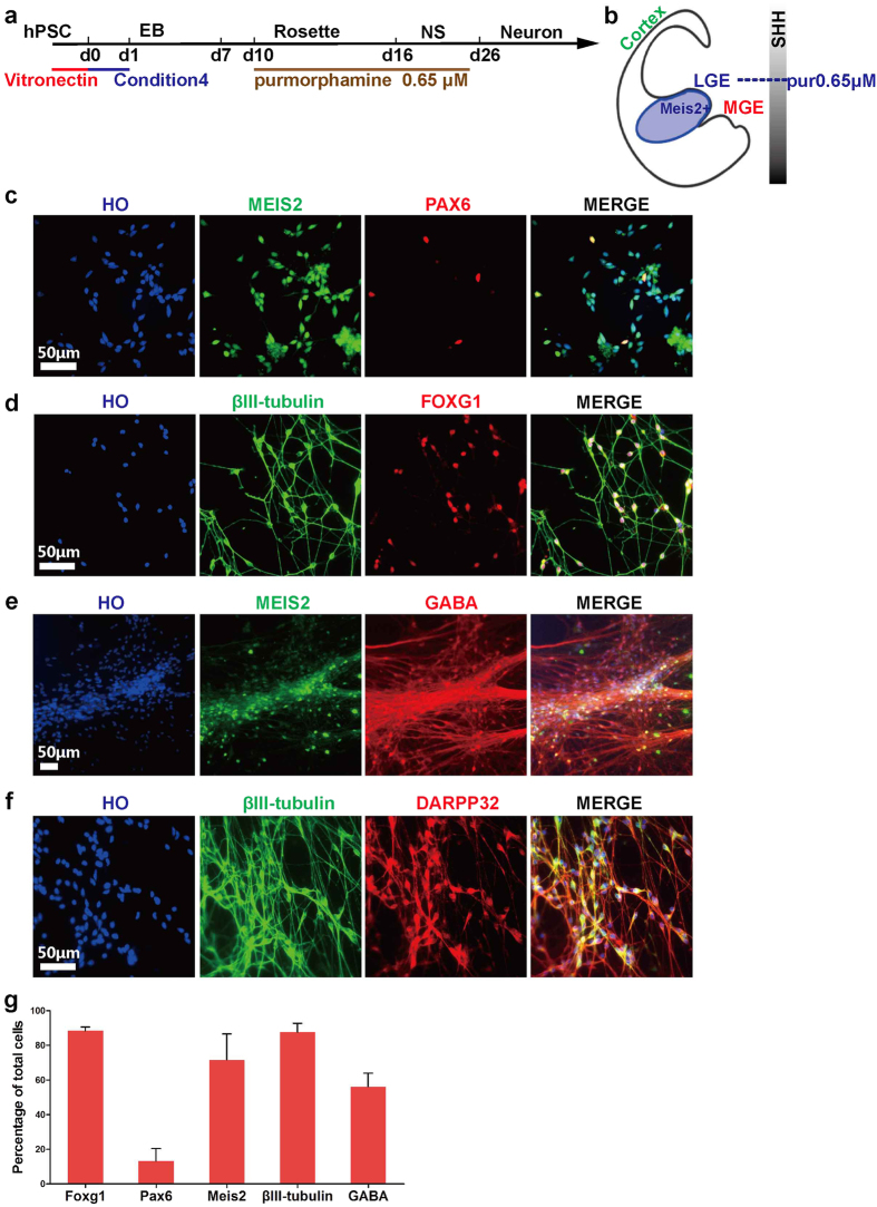
Figure 4. Directed differentiation of hPSCs to striatal medium spiny neurons in a xeno-free system.
(a) Timeline of direct differentiation of hPSCs to striatal medium spiny neurons in a xeno-free system by using optimized condition 4 with low dosage of purmorphamine. (b) A schematic showing coronal section of a human embryo with LGE patterning (blue) containing cells expressing marker MEIS2. (c) At d22, 71.7 ± 3.7% of cells expressed MESI2, but only 13.2 ± 3.0% of cells expressed PAX6, demonstrating cells with LGE fate. Scale bar, 50 μm. (d) At d25, nearly all cells expressed FOXG1 and βIII-tubulin. Scale bar, 50 μm. (e) At d35, GABA + neurons co-expressed MEIS2. Scale bar, 50 μm. (f) At d35, most neurons expressed striatal projection neuron marker DARPP32. Scale bar, 50 μm. (g) Quantification of FOXG1 + , PAX6 + , MEIS2 + , βIII-tubulin + and GABA + cells of total cells.

