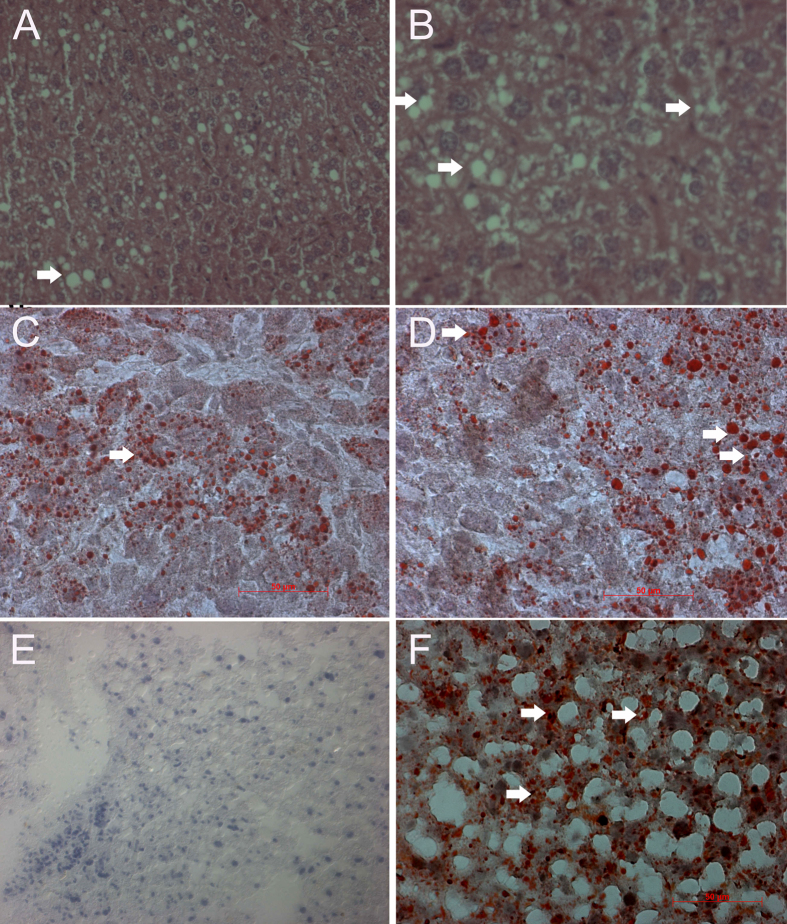
Figure 4. Oil-red ‘O’ staining for pathological examination of liver of α-tocopherol- and β-carotene-treated Apoe−/− mice.
(A,B) Moderate vacoular degeneration (arrow) in the hepatocytes of β-carotene-treated Apoe−/− mice. (C,D) β-carotene-treated liver show moderate accumulation of red colour lipid droplets in the cytoplasm of hepatocytes (arrow). (E) α-tocopherol-treated liver lacking accumulation of lipid droplets as compared to positive control high fat diet-treated liver of Apoe−/− mice. (F) Accumulation of red colour lipid droplets in the cytoplasm of hepatocytes in high fat diet-treated liver of Apoe−/− mice (arrow) (positive control).

