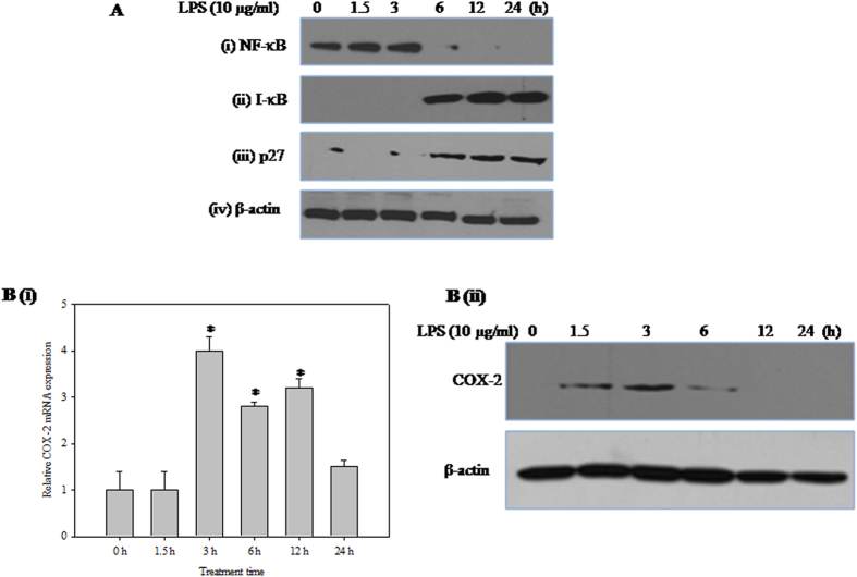
Figure 2. Effect of LPS on corneal epithelial cell signaling.
(A) Effect of LPS on the levels of (i) NF-κB-p65 (ii) I-κBα and (iii) p27 proteins (iv) β-actin was used as an internal control. HCE cells were treated with LPS (10 μg/ml) for different time periods (0, 1.5, 3, 6, 12 and 24 h). Equal amounts of total protein (for I-κB α, p27 and β-actin) and nuclear protein (for NF-κB-p65) was analysed by SDS-PAGE (10–12%), and after electrophoresis under same experimental conditions, proteins on the gel were transferred on to nitrocellulose membrane and probed with specific antibodies. The representative images of three independent experiments shown were cropped. (B) Effect of LPS on the expression of COX-2. (i) Quantitative analysis of COX-2 mRNA expression in HCE cells relative to 18S rRNA expression at different time points (0, 1.5, 3, 6, 12 and 24 h) after LPS treatment by qRT-PCR is reported as fold change relative to 0 h calculated using 2−ΔΔCT. X-axis represents exposure time in hours. Values are mean ± SEM for three independent experiments. *Indicates significance at p < 0.05. (ii) HCE cells were treated with LPS (10 μg/ml) for different time periods (0, 1.5, 3, 6, 12 and 24 h). Equal amounts of protein was analyzed by SDS–PAGE (12%), proteins on the gel were transferred on to nitrocellulose membrane and probed with COX-2 specific antibody under same experimental conditions. The representative images of three independent experiments shown were cropped.

