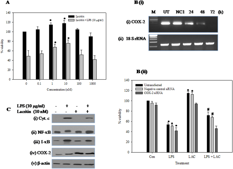
Figure 4. Effect of recombinant lacritin on HCE cells.
(A) Effect of recombinant lacritin on HCE cells with/without LPS. HCE cells were treated with either lacritin alone (0. 0.1, 1, 10, 100, 1000 nM) or lacritin (0, 0.1, 1, 10, 100, 1000 nM) and LPS (10 μg/ml) for 24 h. Viability of untreated control cells was 100% and data is expressed as mean percent of untreated control ± SEM for three independent experiments. *Indicates significant difference at p < 0.05 when compared to untreated control. (B) Effect of COX-2 knockdown on lacritin’s cytoprotection. (i) Semi quantitative RT-PCR analysis of COX-2 expression in the HCE cells transfected or untransfected (UT) with NC1 or COX-2 siRNA for 24, 48 and 72 hours. 18S rRNA served as internal control. (ii) Untransfected or NC1 transfected or COX-2 depleted HCE cells were treated with either LPS (10 μg/ml) alone or lacritin alone (10 nM) or both for 24 h. Viability of untransfected control cells was 100% and data is expressed as mean percent of untreated control ± SEM for three independent experiments. *Indicates significant difference at p < 0.05 when compared to untreated control. #Indicates significant difference at p < 0.05 when compared to their respective LPS alone treated cells. (C) Effect of recombinant lacritin on the levels of (i) Cyt c (ii) NF-κB-p65 (iii) I-κB α and (iv) COX-2 in LPS treated HCE cells. HCE cells were treated with either LPS (10 μg/ml) alone or lacritin (10 nM) alone or both for 24 h. Equal amounts of total protein (for I-κBα and β-actin), nuclear protein (for NF-κB-p65) and cytosolic protein (for Cyt c) was analysed by SDS- PAGE (10–12%), and after electrophoresis under same experimental conditions, proteins on the gel were transferred on to nitrocellulose membrane and probed with protein specific antibodies. The representative images of three independent experiments shown were cropped.

