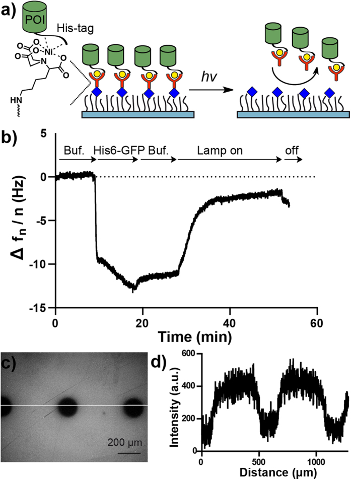
Figure 2. Characterization of NTA-NO2.
(a) Working model of NTA-NO2. The His6-tagged POI (protein of interest) binds to the Ni2+-NTA-NO2 complex presented at the surface. Upon illumination the NTA group is cleaved from the surface and the protein is released. (b) His6-GFP binds on SiO2 QCM crystals coated with 10 mol% PEG-azide and modified with NTA-NO2. When the lamp is turned on the His6-GFP is liberated from the surface. (c) Fluorescence image of NTA-NO2 modified surface with circular micropatterns (160 μm) and (d) the line profile along the line. The surface was illuminated under an upright fluorescence microscope with an adjustable field aperture. The surface was incubated afterwards with Ni2+ and His6-GFP for visualization.

