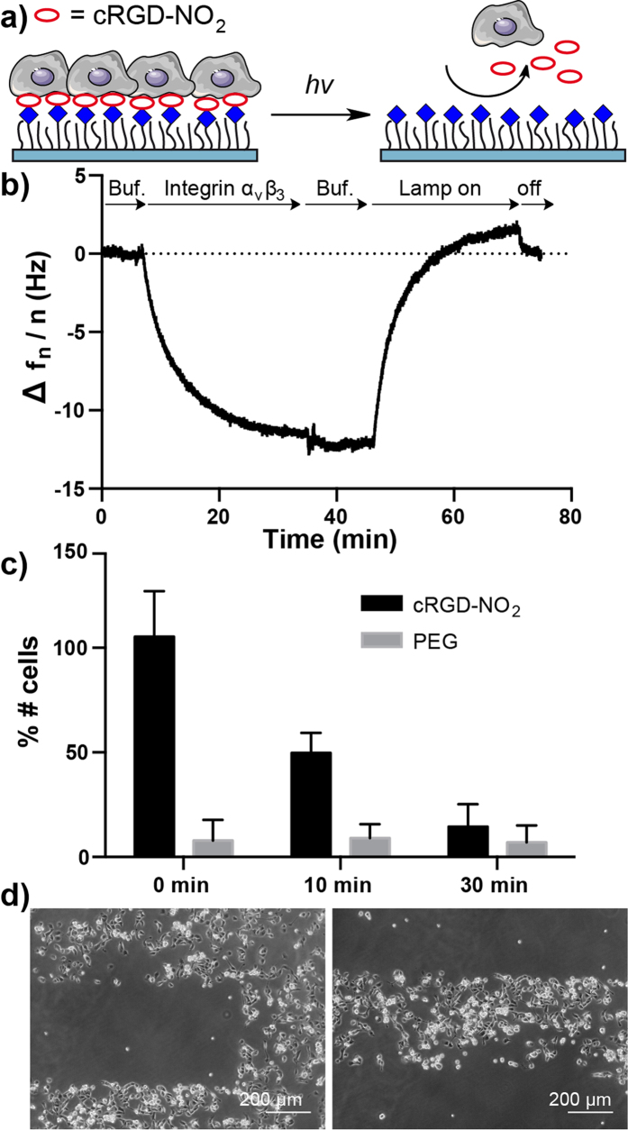
Figure 3. Characterization of cRGD-NO2.
(a) Working model of cRGD-NO2. Cells can adhere to cRGD-NO2 modified PEG surfaces through integrin binding. Upon illumination the integrin ligand cRGD is cleaved off and cells can no longer adhere on the PEG coating. (b) QCM measurements showing integrin αvβ3 binding to cRGD-NO2 modified 1 mol% PEG-azide surfaces. The bound integrin αvβ3 is washed off as the surface is irradiated (λ = 365 nm). (c) REF cell adhesion on surfaces with 1% PEG-azide coating modified with cRGD-NO2. Surfaces are irradiated with light of λ = 365 nm for 0, 10 and 30 minutes to achieve various extends of cRGD cleavage. Unmodified surfaces with the same PEG coating are used as controls and the number of cells on the surface are given as the percentage of cells that adheres on a cRGD modified surface. The error bars are the standard deviation from three independent experiments. (d) Patterned MDCK cells on cRGD-NO2 modified surfaces. The surface was illuminated under an upright fluorescence microscope with an adjustable field aperture.

