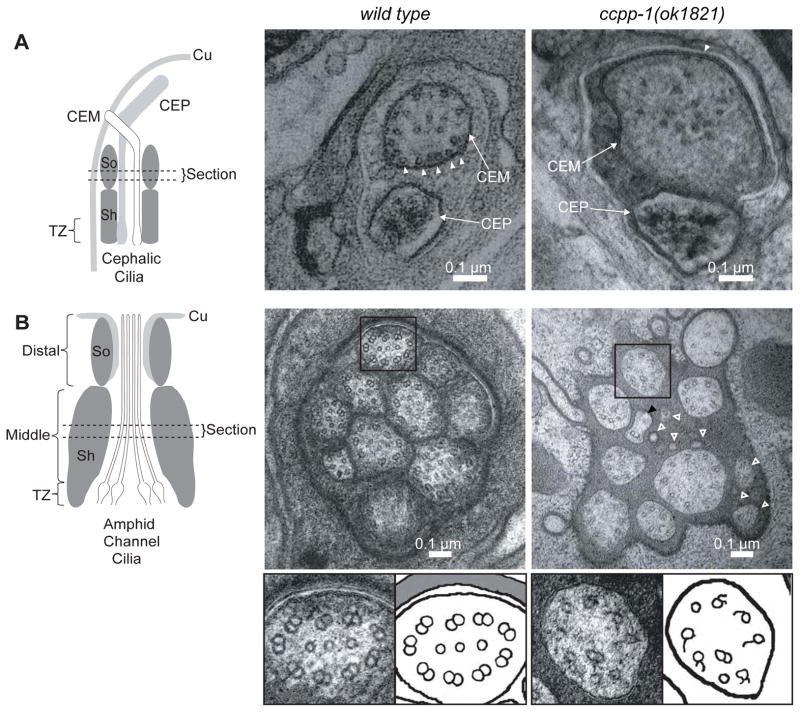
Fig. 3. ccpp-1(ok1821) mutants exhibit ciliary ultrastructure defects.
Left diagrams show regions from which the cephalic (CEM and CEP) and amphid cilia images were taken (Cu = cuticle; Sh = sheath cell; So = socket cell; TZ =transition zone). A EM Images of CEM and CEP cilia in wild-type and ccpp-1(ok1821) mutant adult males. Wild-type CEM cilia image (taken from a tomogram) contained many singlet MTs (19 singlets in section shown) closely apposed to the membrane (arrowheads). ok1821 CEM cilia had fewer singlets (15 singlets in section shown), which were more distant from the membrane (arrowhead indicates one singlet near membrane). The ok1821 CEM cilium diameter was larger than wild type. B Thin sections of amphid cilia in wild-type and ccpp-1(ok1821) adult males. Wild-type middle segments contain ten axonemes, each of which typically has nine outer doublets plus a variable number of inner singlets. The ok1821 middle segment contained only eight intact axonemes plus what appear to be fragments of two cilia (hollow white arrowheads), one of which contains a singlet with attached broken B-tubule (black arrowhead). Most ok1821 mutant axonemes had fewer MTs, with many doublets replaced by singlets or broken B-tubules. Bottom, boxed wild-type and mutant axonemes accompanied by cartoons. Refer to Table 1 for quantification of images.

