Fig. 3.

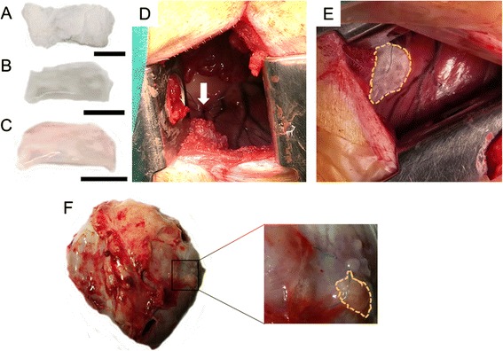
Engraftment of a decellularized myocardial ECM scaffold embedded with cells in a swine myocardial infarction model. a Lyophilized and gamma ultraviolet sterilized decellularized myocardial ECM scaffold. b, c Decellularized scaffold after the addition of peptide hydrogel (b) and porcine adipose tissue-derived progenitor cells (c). Scale bars = 1 cm. d Image of the myocardial infarction site, induced by double ligation in the first marginal branch of the circumflex artery (indicated with white arrow). e Reseeded decellularized scaffold placed over the injured myocardium. Scaffold is indicated with yellow dotted lines. f Presence of the implanted scaffold on the infarcted area in explanted hearts 28 days after sacrifice. The remaining scaffold is highlighted with yellow dotted lines
