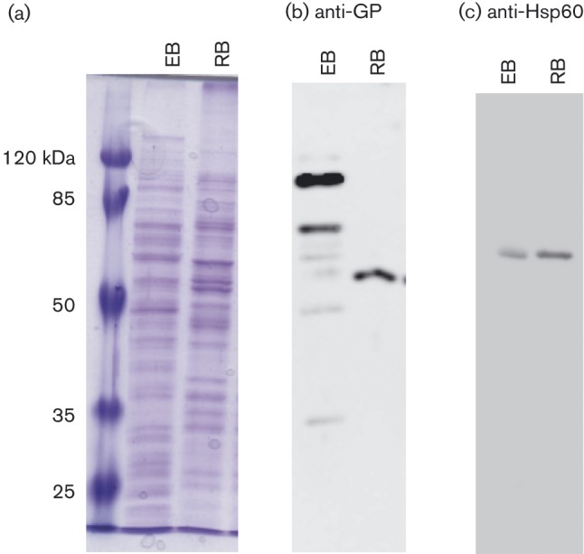
Fig. 1. 1D SDS-PAGE and Western blot analysis of C. caviae EBs and RBs. EB and RB samples were lysed using 2D lysis conditions and proteins were methanol-precipitated for 1D SDS-PAGE analysis. Samples were run on 12 % SDS-PAGE and then either stained with Coomassie brilliant blue (a) or transferred to nitrocellulose membranes for Western blot analysis (b and c). Membranes were probed either with crude antisera from C. caviae-infected guinea pigs (b) or with monoclonal anti-Hsp60 antibody A57-B9 (c) followed by horseradish peroxidase-conjugated anti-guinea pig or anti-mouse IgG antibodies, respectively. The protein sample source (EB or RB) is listed at the top of each panel. Molecular mass markers are shown to the left of (a).

