. 2015 Aug;96(Pt 8):2104–2113. doi: 10.1099/vir.0.000178

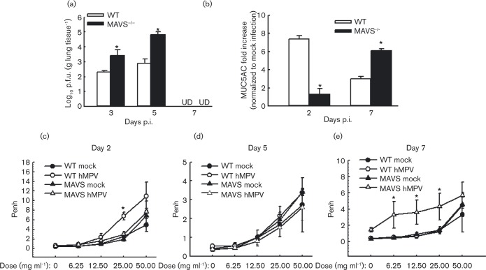
Fig. 5. Effect of MAVS on lung viral replication and function. WT and MAVS− / −  mice were infected i.n. with 1 × 107 p.f.u. of hMPV or mock-infected, total lung was harvested at indicated time points p.i. and lung infectious viral particles were titrated on LLC-MK2 cell monolayers by immune staining assays (a) and MUC5AC was quantified by real-time PCR (b). (c–e) Lung function in hMPV-infected mice, either WT or MAVS, at days 2, 5 and 7 p.i. was assayed by post-methacholine challenge. Penh values were determined by unrestrained plethysmography. n = 3–5 mice in each group. The results are from two independent experiments. *P < 0.05 for comparison with hMPV-infected WT or MAVS− / −  mice. UD, undetectable.

Fig. 5.