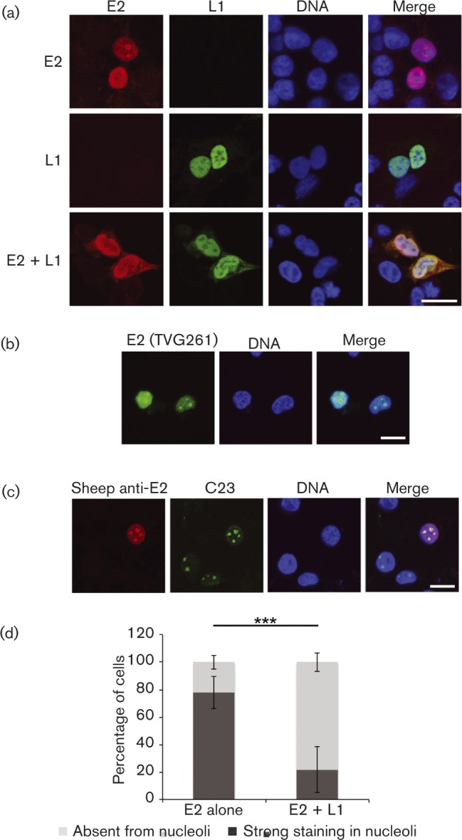
Fig. 5. Subcellular localization of HPV16 E2 and L1. Confocal imaging. (a) C33a cells transfected with E2 and L1 expression plasmids alone or in combination. E2 protein was stained with sheep anti-E2 antibody (red) and L1 protein was stained with mouse anti-L1 antibody (green). DNA was stained with Hoechst (blue). (b) E2 protein was detected with mouse monoclonal anti-E2 antibody (TVG261; green). (c) E2 protein was detected with sheep anti-E2 antibody (red), and nucleolar marker protein C23 is shown in green. Bars (all panels), 10 μm. (d) E2-expressing cells transfected alone or in combination with L1 were scored for strong E2-staining in the nucleolus or E2-staining absent from the nucleolus. At least 80 cells were scored for each experiment. The data shown represent the mean ± se of three independent experiments. Significance was determined using Student's t-test; ***P < 0.001.

