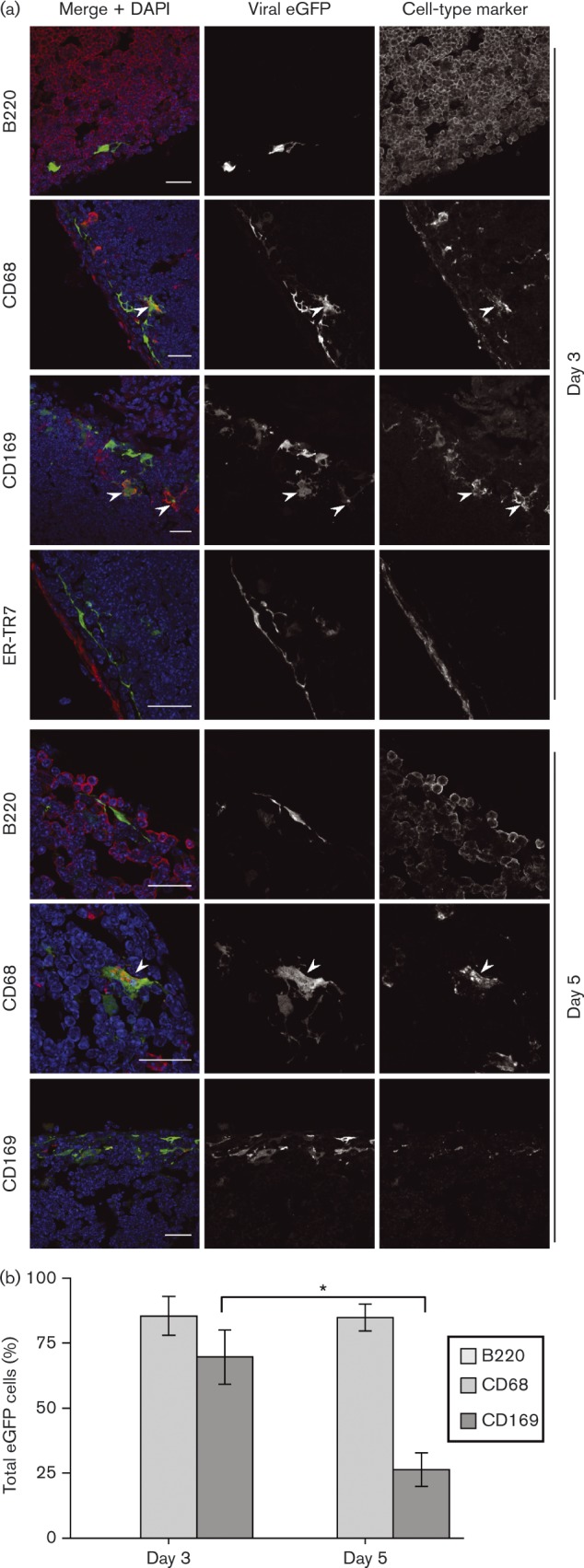
Fig. 3. PLN infection by i.f. replication-deficient MuHV-4. (a) C57BL/6 mice were infected i.f. with replication-deficient MHV-GFP (ORF50− ). Sections of PLN harvested 3 and 5 days later were stained for viral eGFP (green in merge) and cell-type markers (red in merge). Nuclei were stained with DAPI (blue). The images are representative of six mice per time point. Arrows show examples of green/red co-localization. Scale bar = 10 μm. (b) EGFP+ cells were counted for five mice (three sections per mouse). Bars show mean ± sem for eGFP+ cells expressing each marker (co-localization with ER-TR7 was < 2 %). From day 3 to day 5, CD169+eGFP+ cell numbers decreased significantly (*P < 0.02). This was associated with a general loss of CD169 staining, seen also with wild-type infection.

