. 2015 Aug;96(Pt 8):2314–2327. doi: 10.1099/vir.0.000140

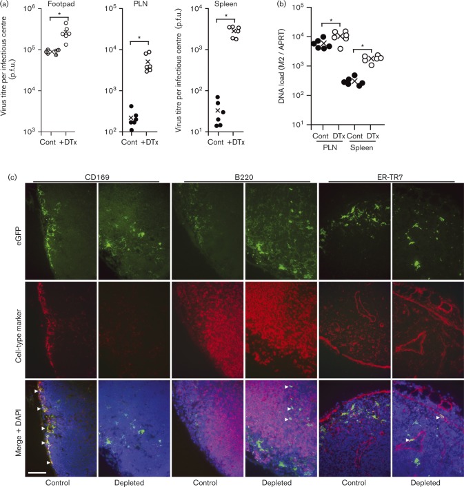
Fig. 8. Effect of CD169+ cell depletion on the spread of i.f. MuHV-4. (a) CD169-DTR mice were depleted or not of CD169+ cells by i.p. DTx injection and then infected i.f. with MHV-GFP. Three days later, virus was titrated in footpads by plaque assay and in PLNs and spleens by infectious centre assay. ○, ⊕ and •, Individual mice; × , means. DTx treatment significantly increased virus titres in each site (*P < 0.01), with the increase in splenic titres being significantly greater than in footpads or PLN (P < 0.01). (b) Mice were depleted or not and then infected as in (a). Three days later, viral DNA loads were assayed in PLNs and spleens by PCR (M2 gene) and normalized by the cellular DNA load of each sample, assayed in parallel (APRT gene). DTx treatment significantly increased viral DNA loads in PLNs (**P < 0.05) and spleens (*P < 0.01). (c) Mice were depleted or not and then infected as in (a). Three days later, PLN sections were stained for virus-expressed eGFP (green) and for cell-type-specific markers (red). Nuclei were stained with DAPI (blue). Arrows show examples of green/red co-localization. Bar, 50 μm.

Fig. 8.