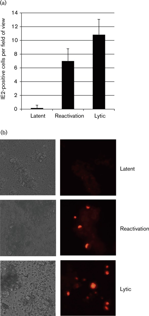
Fig. 2. Latent infection is established in Kasumi-3 cells at 3 days post-infection. Undifferentiated Kasumi-3 cells were infected with IE2–RFP tagged HCMV TB40E strain at an m.o.i. of 3.0 for 3 days before being analysed (latent) or following the establishment of latency for 3 days. Kasumi-3 cells were treated with phorbol 12-myristate 13-acetate as described previously (O'Connor & Murphy, 2012) for 48 h before analysis (reactivation). Finally, prior to infection, cells were differentiated with phorbol 12-myristate 13-acetate for 48 h (lytic). Cells were counted and presented graphically (a) and were analysed directly by immunofluorescence (b). Data in (a) represent triplicate samples of six fields of view (means ± sd).

