. 2015 Aug;96(Pt 8):2453–2463. doi: 10.1099/vir.0.000169

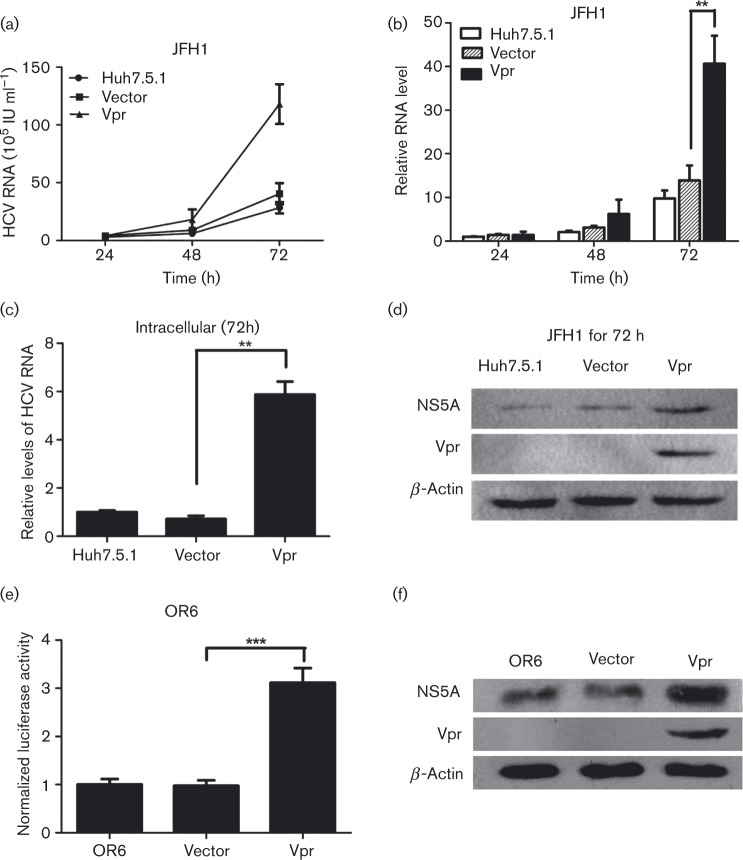
Fig. 1. Vpr stimulates HCV replication in JFH1 infection model and OR6 cell lines. (a) Quantitative HCV RNA assay in cell culture supernatants from Huh7.5.1 cells transfected with Vpr and then infected with JFH1 at different times. (b) The relative fold change of HCV RNA levels in cell culture supernatants from Huh7.5.1 cells was measured, with the control non-transfected JFH1-infected Huh7.5.1 cells at 24 h being assigned a value of 1. (c) Quantitative PCR for intracellular HCV RNA levels in the transient Vpr or vector-transfected Huh7.5.1 cells at 72 h post-JFH1 infection. (d) Western blotting confirmed the expression of β-actin, NS5A and Vpr in Huh7.5.1 cell lines infected with JFH1. (e) Renilla luciferase assay in OR6 cell lines transfected with Vpr and controls. (f) Western blot analysis in OR6 cell lines confirmed the expression of β-actin, NS5A and Vpr. The data are presented as mean ± sd. **P < 0.01, ***P < 0.001.

Fig. 1.