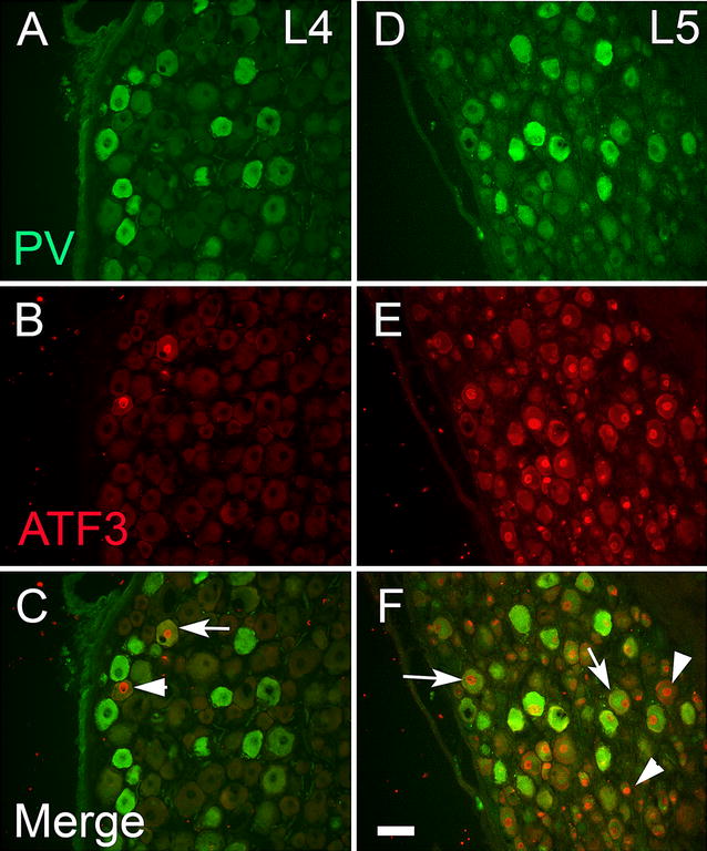
Fig. 3.

Parvalbumin (green) and ATF3 (red) staining in the ipsilateral L4 (A, C, E) and L5 (B, D, F) ganglia 2 weeks after L5 spinal nerve ligation and section. Arrows denote examples of double-labelled cells; arrowheads denote examples of cells that only express ATF3. Almost all PV-ir cells were ATF3-ir in the L5 ganglion (arrows). Very few ATF3 cells were seen in L4 DRG. Scale bar 75 μm
