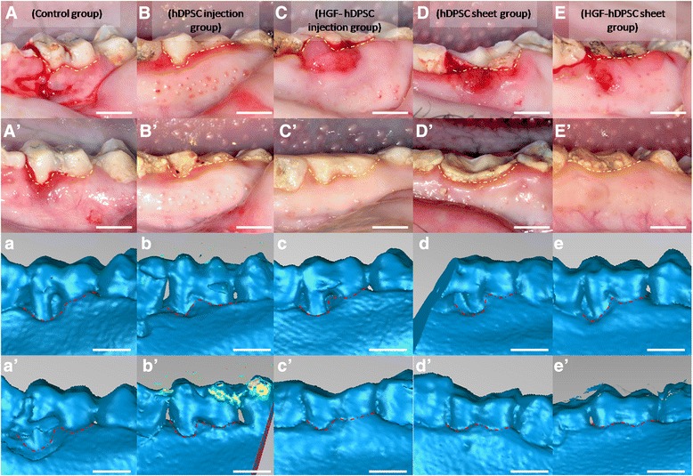
Fig. 4.

Regeneration of tissue in periodontal defects mediated by transplanted hDPSCs. Intraoral photographs were acquired A–E before transplantation or A′–E′ 12 weeks after transplantation. A′ Only limited periodontal tissues were regenerated in the control group (yellow dotted line). Marked periodontal tissue formation was found in B′ the hDPSC injection group and C′ the HGF-hDPSC injection group, but they did not restore tissues to healthy levels (yellow dotted line). Periodontal tissue regeneration mediated by D′ the hDPSC sheet and E′ the HGF-hDPSC sheet achieved close to normal tissue levels (yellow dotted line). Three-dimensional CT images were acquired a–e before transplantation and a′–e′ 12 weeks after transplantation. a′ Only limited bone regeneration was observed in the control group, but marked bone formation was observed b′ in the hDPSC injection group, c′ the HGF-hDPSC injection group, d′ the hDPSC sheet group, and e′ the HGF-hDPSC sheet group after cell transplantation (red dotted line). Yellow dotted line, gingival margin; red dotted line, bone margin. hDPSC human dental pulp stem cell, HGF hepatocyte growth factor
