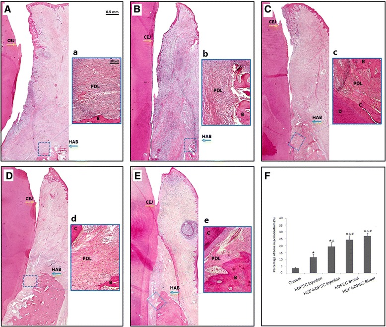
Fig. 6.

Whole-view histopathological assessments of periodontal tissue regeneration in H & E stained sections. New periodontal tissue regeneration within the periodontal defect is shown for the A control group, B hDPSC injection group, C HGF-hDPSC injection group, D hDPSC sheet group, and E HGF-hDPSC sheet group. New bone, cementum, and periodontal ligament were regenerated in the periodontal defect areas that received a no treatment, or treatments of b hDPSC injections, c HGF-hDPSC injections, d hDPSC sheets, or e HGF-hDPSC sheets. Alveolar bone regeneration in A the control group was much less pronounced than that observed in the C HGF-hDPSC injection group, D hDPSC sheet group, and E HGF-hDPSC sheet group. Newly formed Sharpey’s fibers were observed penetrating into the newly regenerated cementum in the c HGF-hDPSC injection group, d hDPSC sheet group, and e HGF-hDPSC sheet group. F The percentages of periodontal bone in the hDPSC injection, HGF-hDPSC injection, hDPSC sheet, and HGF-hDPSC sheet groups were significantly higher than that of the control group (*P <0.01). The percentages of periodontal bone were higher in the HGF-hDPSC injection, hDPSC sheet, and HGF-hDPSC sheet groups than in the hDPSC injection group (Δ P <0.01). The percentages of periodontal bone were higher in the hDPSC sheet and HGF-hDPSC sheet groups than in the HGF-hDPSC injection group (# P <0.01). B bone, C cementum, CEJ, cementum–enamel junction, D dentin, HAB height of alveolar bone, hDPSC human dental pulp stem cell, HGF hepatocyte growth factor, PDL periodontal ligament
