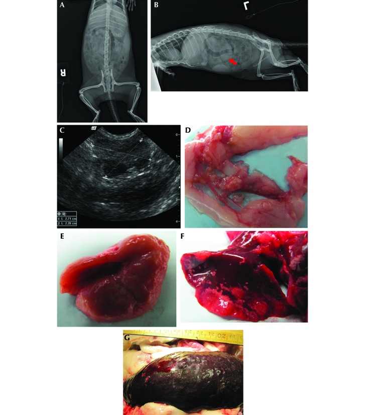
Figure 3.
Case 3. (A) Ventrodorsal and (B) left lateral radiographs. Radiopaque foreign material is present in both views. The lateral view shows a poorly defined midabdominal area of increased density (arrow). (C) Ultrasonographic image of the midabdominal mass 5 wk after abdominal exploratory. (D) Multiple, firm, tan, 1- to 5-mm nodules throughout the mesentery. (E) Pitting lesions on kidney. (F) Multifocal, coalescing, raised white nodules on the lungs. (G) Enlarged spleen with mulitfocal pitting lesions.

