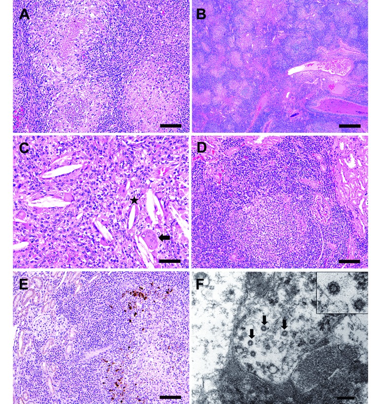
Figure 4.
Case 3. (A) Multifocal to coalescing pyogranulomatous and lymphoplasmacytic lymphadenitis and peritonitis, with histologic features of central necrotic cores surrounded by neutrophils, macrophages and lymphocytes (bar, 100 µM). (B) Severe granulomatous and lymphoplasmacytic pneumonia, with pulmonary architectural effacement and consolidation. Discernable bronchial and bronchiolar lumens are filled with eosinophilic proteinaceous material, mucus, and epithelial and inflammatory cellular debris (bar, 500 µM). (C) Needle-like acicular cholesterol clefts (*) that are often associated closely with multinucleated giant cells (arrow; bar, 50 µM). (D) Cortical lymphoplasmacytic interstitial nephritis (bar, 100 µM). Hematoxylin and eosin stain (A through D) (E) Positive staining for feline coronavirus (FIPV3-70) monoclonal antibody within macrophages in granulomatous foci in the kidney, whereas plasma cells and lymphocytes in the interstitium are negative (bar, 100 µM). (F) Electron microscopy of one of the mesenteric nodules reveals intracellular coronavirus-like particles (arrows). Virions were present in several nodules selected for examination and were found both within vacuoles and free in cytoplasm. Inset shows higher resolution image of a viral particle with the characteristic radiating crown-like coronavirus morphology (bar, 200 nM).

