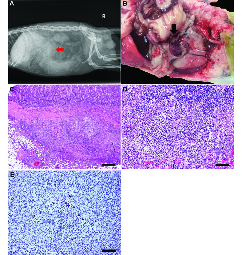
Figure 5.
Case 5. (A) Right lateral radiograph depicting splenomegaly, decreased serosal detail, and an area of increased opacity (arrow) in the cranial midabdomen. (B) Nodular mesenteric mass firmly adhered to the small intestine (arrow). (C) Pyogranulomatous inflammation disrupts and effaces mesentery and small intestine. Hematoxylin and eosin stain; bar, 500 µm. (D) Pyogranulomatous inflammation disrupts hepatic architecture. Inflammation is composed of minimal number of degenerate neutrophils surrounded by moderate numbers of epithelioid macrophages, lymphocytes, plasma cells, and fewer fibroblasts. Hepatocytes adjacent to the affected area have intracytoplasmic vacuoles (lipid type). Hematoxylin and eosin stain; bar, 100 µm. (E) Epithelioid macrophages surrounding core neutrophils of a pyogranulomatous focus reveal strong intracytoplasmic immunoreactivity to feline coronavirus (FIPV3-70) monoclonal antibody (bar, 100 µm).

