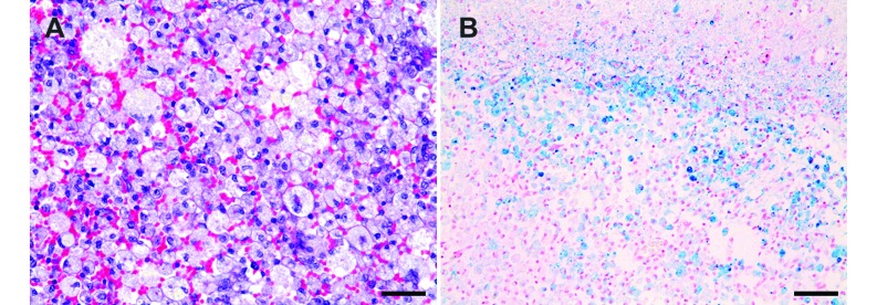
Figure 5.
Microscopic features of the canine brain after experimentally induced ICH. (A) Cavitated areas containing numerous macrophages and extravasated erythrocytes were identified in the white matter of the parietal lobe. Hematoxylin and eosin stain; magnification, 400×; Scale bar, 35 µm. (B) Prussian blue stain was found in macrophages, which were distributed among discrete lesions of white matter. Magnification, 200×; Scale bar, 70 µm.

