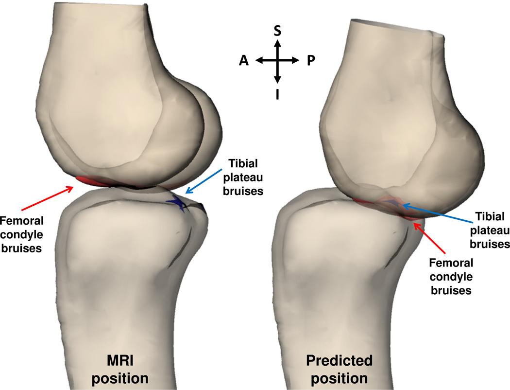
Figure 2.
Numerical optimization was used to maximize overlap of bone bruises on the femoral condyle (red) and tibial plateau (blue) and predict the position of injury. A sagittal view of a 3D model of one subject’s knee is shown in the MRI position (left) and in the predicted position of injury (right). (A=anterior, P=posterior, S=superior, I=inferior)

