
Figure 8.
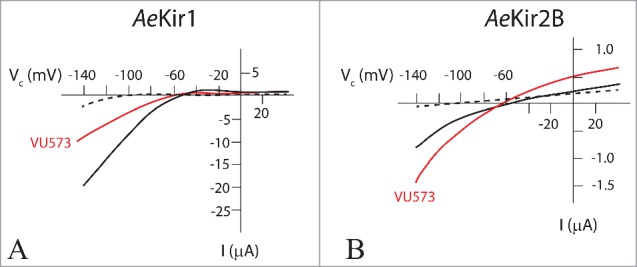
Effect of VU573 on AeKir and AeKir2B expressed in Xenopus oocytes. Oocytes were bathed in Ringer solution containing 0.5 mM K+ (broken line), and 10 mM K+ (solid lines) in the absence (black) and presence (red) of VU573 (50 µM). Note the different current scales in A and B. VU573 inhibits AeKir1 (A) and stimulates AeKir2b (B). Data adapted from Rouhier et al.54