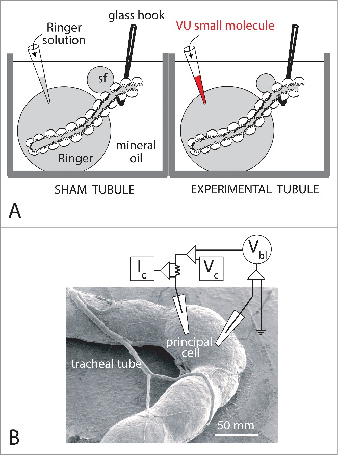
Figure 9.

Methods for studying Malpighian tubules in vitro. A. The modified Ramsay fluid secretion assay.78 “Sister tubules” isolated from the same female yellow fever mosquito are studied in parallel. The blind, secretory end of each tubule is bathed in a Ringer droplet (50 µl) under light mineral oil. The open end of the tubule is pulled into mineral oil. A nick in the epithelium allows fluid secreted by the tubule to escape the lumen as a droplet separate from the Ringer bath. To begin, both tubules are first studied for a 30 min control period. Thereafter, 5 µl of Ringer solution is replaced with an equal volume of Ringer solution plus 1) 0.5 % DMSO (vehicle) in the sham tubule, and 2) 0.5% DMSO and 100 µl VU small molecule. Secreted fluid was collected for the analysis of its composition by electron probe.79,80 B. TECV studies of a principal cell of an Aedes Malpighian tubule.61 Both voltage and current microelectrodes impale a single principal cell of the tubule. Vbl, basolateral membrane voltage; Vc, voltage command; Ic, clamp current. Stellate cells are too thin (<10 µM) for stable measurements with intracellular microelectrodes. Data from Wu et al.81 and Hine et al.9
