
Fig. 1. UL138 inhibits VPA-responsive IE1 expression during HCMV latency.
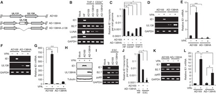
(A) Schematic of recombinant AD169 viruses encoding UL138 (see also fig. S2A). (B) RNA from THP-1 and primary CD34+ cells infected at a multiplicity of infection (MOI) of 1 with the indicated viruses for 18 or 24 hours, respectively, was analyzed for transcript expression of the indicated gene by reverse transcription polymerase chain reaction (RT-PCR). RNA from normal human dermal fibroblasts (NHDFs) infected with AD-138HA at an MOI of 1 for 24 hours serves as a positive control. (C) RNA and DNA from samples in (B) were analyzed by quantitative RT-PCR (qRT-PCR) or qPCR, respectively, and normalized to cellular glyceraldehyde-3-phosphate dehydrogenase (GAPDH). Viral transcripts were normalized to viral DNA and are shown relative to that of NHDFs (HF). (D and E) CD34+ cells infected at an MOI of 1 for 24 hours in the absence (−) or presence (+) of VPA (1 mM) analyzed for transcript expression by RT-PCR (D) or qRT-PCR (E). (F to H) THP-1 cells infected at an MOI of 3 for 18 hours in the absence (−) or presence (+) of VPA analyzed for transcript expression by RT-PCR (F) or qRT-PCR (G) and protein expression by Western blot (H). (I) ESCs infected with the indicated virus at an MOI of 3 for 24 hours analyzed for transcript expression of the indicated gene by RT-PCR. (J) ESCs infected as in (I) were analyzed for IE1 transcripts per viral genome as in (C). (K) ESCs infected with the indicated virus at an MOI of 3 for 24 hours in the absence (−) or presence (+) of VPA analyzed for transcript expression of the indicated gene by RT-PCR. (L) Samples from (K) were analyzed by qRT-PCR. Data are means ± SEM from at least three independent experiments. *P < 0.05, **P < 0.01, or ***P < 0.001 by Student’s t test.