
Fig. 2. UL138 is not required for HDAC-independent repression of IE gene expression in clinical strains and is not sufficient to maintain latency.
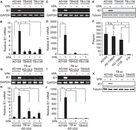
(A to D) CD34+ (A and B) or THP-1 (C and D) cells infected with AD169, TB40/E, or TB40/E-ΔUL138 (TB-Δ138) at an MOI of 1 in the absence (−) or presence (+) of VPA were analyzed for the indicated transcripts by RT-PCR (A and C) or qRT-PCR (B and D). (E) Lysates from THP-1 cells infected as in (C) analyzed by Western blot. (F) Infectious virions produced by ESCs infected with the indicated virus at an MOI of 3 for 10 days were quantitated by plaque assay. (G to J) CD34+ (G and H) or THP-1 (I and J) cells infected with AD169, TB40/E with ULb′ replaced with that of AD169 (TB w/AD-ULb′), or TB40/E at an MOI of 1 in the absence (−) or presence (+) of VPA were analyzed by RT-PCR (G and I) or qRT-PCR (H and J). (K) Lysates from THP-1 cells infected as in (I) analyzed by Western blot. Data are means ± SEM from three independent experiments. *P < 0.05 or **P < 0.01 by Student’s t test. n.s., not significant (P = 0.29).