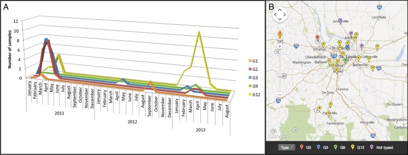
Figure 2.
Rotavirus VP7 serotypes (G types) 2011–2013. (A) Samples determined to be positive for rotavirus (Fig. 1) were typed using a targeted polymerase chain reaction (PCR) and amplicon sequencing approach. Different colored lines represent each subtype detected. The number of samples of each subtype (y-axis) is plotted over time (x-axis). (B) The G12 serotype was distributed throughout the St. Louis metropolitan area in the 2012–2013 rotavirus season. Zip codes were used to map the cities of residence of the patients from the 2012–2013 rotavirus season. This includes patients from St. Louis Children's Hospital, Mercy Hospital, and Cardinal Glennon Children's Medical Center. Not shown are 3 patients from more remote locations in Missouri and Illinois (2 G12, 1 G2) and 1 patient from Arkansas (G12). Note that “not typed” indicates that the reverse transcriptase–PCR/sequencing reactions failed.

