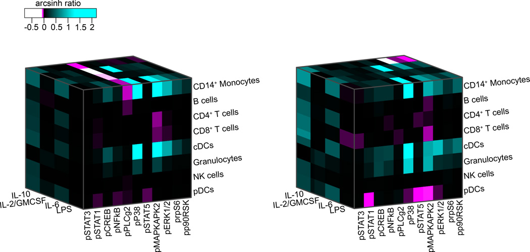
Figure 4. Patient-specific evoked signaling responses in myeloid cell subsets.
Patient-specific immune states before surgery were characterized along three axes including eight cell types, eleven signaling proteins, and the activity of these signaling proteins in response to four different extracellular ligands. Signaling activity is indicated by the color-coded arcsinh ratio, i.e., the difference between arcsinh transformed values (inverse hyperbolic sine) obtained in unstimulated and stimulated conditions. A total of 352 parameters described each patient’s immune state. Shown are the three-dimensional heat maps characterizing and differentiating the immune states of two exemplary patients. Abbreviations: IL-10, interleukin 10; IL-2, interleukin 2; GMCSF, granulocyte monocyte colony-stimulating factor; IL-6, interleukin 6; LPS, lipopolysaccharide; pSTAT1/3/5, phosphorylated signal transducer and activator of transcription 1/3/5; pCREB, phosphorylated cAMP response element-binding protein; pNFkB, phosphorylated nuclear factor kappa-light-chain-enhancer of activated B cells; pPLCg2, phosphorylated phospholipase C gamma 2; pP38, phosphorylated P38 map kinase; pMAPKAPK2, phosphorylated mitogen activated protein kinase-activated protein kinase-2; pERK1/2, phosphorylated extracellular regulated kinase 1/2; prpS6, phosphorylated ribosomal protein S6; pp90RSK, phosphorylated ribosomal S6 kinase; pDCs, plasmacytoid dendritic cells; cDCs, classical dendritic cells.

