Figure 6.
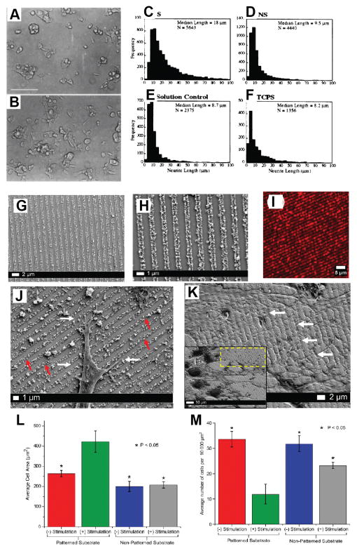
PC-12 cell differentiation on PPy without (A) and with (B) application of an electric potential. PC-12 cells were grown on PPy for 24 h in the presence of NGF, then exposed to electrical stimulation (100 mV) across the polymer film, S (B). Images were acquired 24 h after stimulation. Cells grown for 48 h but not subjected to electrical stimulation, NS, are shown for comparison (A). Bar= 100 μm. Neurite length histograms. Shown are histograms of neurite lengths for cells on PPy with (S, stimulated) (C) and without (NS, unstimulated) (D) potential applied through PPy, on PPy with potential applied through the solution (E), and on tissue culture polystyrene (TCPS) (F). Reproduced with permission.[45] Copyright 1999. Characterization of the multilayered PEDOT:PSS conducting nanoparticles and their assembly as linear conduits. (G, H) SEM micrographs of the nanoparticle patterns at a magnification of 25kX (G) and 11kX (H) indicating the formation of tightly packed and highly ordered nanoparticle arrays. (I) Confocal fluorescence image of the RhB functionalized PEDOT:PSS nanoparticle arrays at 20X magnification. (J) High magnification (12kX magnification) SEM images demonstrating specific and preferential interactions of neurites (white arrows) with the PEDOT:PSS linear conduits (red arrows). (K) High-magnification SEM image (magnification 8kX) indicating the formation of extensive dendritic networks (white arrows) guided by the PEDOT:PSS arrays. Inset: The corresponding low-magnification image of the area (yellow box) analyzed (magnification 3kX). (L) Significant increase in the average cell area is observed 72 h after exogenous electrical stimulation on the nanoparticle platform in comparison to unstimulated and non-patterned controls. (M) Corresponding decrease in PC12 cell proliferation observed 72 h after exogenous electrical stimulation on the nanoparticle platform in comparison to unstimulated and non-patterned controls. Reproduced with permission.[136] Copyright 2015.

