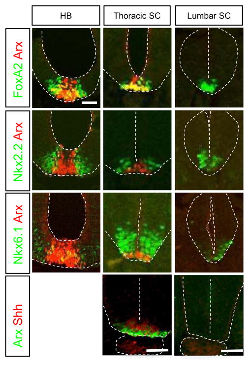
Fig. 1.
Cross-sections of embryonic chick neural tube (HH10) showing Arx, Nkx2.2, Nkx6.1, FoxA2 and Shh expression in the hindbrain (HB) and spinal cord (SC). The colored letters on the left side box indicate the antibodies used for immunohistochemistry. HB (hindbrain), thoracic SC, and lumber SC indicate the level of each section. Scale bar in the upper left panel is 50 μm for all images.

