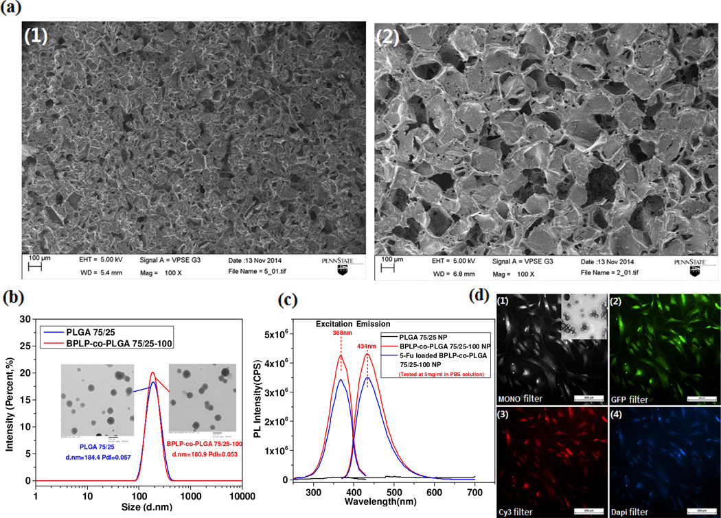
Fig. 6.
Scaffold and nanoparticle fabrication of BPLP-co-PLGA. (a) SEM images of BPLP-co-PLGA 75/25-100 scaffolds with pore sizes of 50-100 µm (1), and 200-250 µm (2). (b) The particle sizes and distributions of representative PLGA and BPLP-co-PLGA nanoparticle dispersions (insert: TEM images of corresponding nanoparticles). (c) Excitation and emission spectra of BPLP-co-PLGA 75/25-100 nanoparticles, 5-Fu loaded BPLP-co-PLGA 75/25-100 nanoparticles and control PLGA 75/25 nanoparticles with a concentration of 5 mg/mL in PBS solution. (d) BPLP-co-PLGA 75/25-50 nanoparticle (500µg/mL)-uptaken hMSCs observed under fluorescence microscope with (1) Monochrome filter (insert, TEM image of BPLP-co-PLGA 75/25-50 nanoparticles, particle size: 110nm), (2) GFP filter, (3) Cy3 filter, and (4) DAPI filter.

