
Figure 3. Characterization of encapsulated cardiomyocytes.
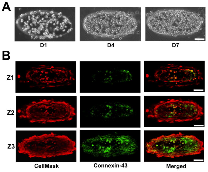
(A) Cell density within the GelMA structures increased as a function of culture time. (B) X-Y confocal sections of cells ubiquitously stained with CellMask and immunostained for Connexin-43. Connexin-43 negative staining amongst the encapsulated cells suggests the presence of cardiac fibroblasts. The Confocal sections proceed from the top, Z1, to the bottom, Z3, of the GelMA structures. Scale bar: 100 μm.