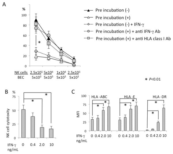
Figure 4.
IFN-γ and HLA class I modulated NK cell cytotoxicity against BEC. BEC were mixed with activated autologous NK cells at an NK/BEC ratio of 0.5 and incubated for 12 hours during pre-incubation (+). After pre-incubation, activated NK cells were added to the co-cultures, in triplicate, to bring the NK/BEC ratio to the indicated levels for a standard 51Cr release assay, in the presence or absence of anti-IFN-γ Ab or anti-HLA class I Ab. BEC without NK cell pre-incubation (−) were also used as target cells with or without addition of IFN-γ. Results are presented as mean +/− S.D. *, significant difference (p<0.01) between pre-incubation (+) versus pre-incubation (−), pre-incubation (−) versus pre-incubation (−) + IFN-γ, pre-incubation (+) versus pre-incubation (+) + anti-IFN-γ Ab, or pre-incubation (+) versus pre-incubation (+) + anti-HLA Ab. B. BEC not pre-incubated with NK cells were co-cultured with autologous activated NK cells at an NK/BEC ratio of 50 for a standard 51Cr release assay in the presence of IFN-γ at the indicated concentrations. Results are presented as mean +/− S.D. *, significant differences (p<0.01) between IFN-γ 0 versus 2.0 or 10. C. BEC not pre-incubated with NK cells were co-cultured for 24 hours in the presence of IFN-γ at the indicated concentrations, then analyzed for the expression of MHC class I and class II molecules by flow cytometry. Results are presented as mean +/− S.D. *, significant differences (p<0.01) between IFN-γ 0 versus 2.0 or 10.

