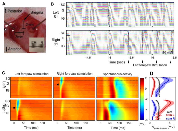
Fig. 1.
Electrophysiological recordings from rat bilateral S1fl using laminar electrodes (n = 6). Two laminar electrodes were located on the forelimb regions of bilateral rat S1 cortices (a). Robust spontaneous activity was observed during rest and forelimb stimulation as shown in example of laminar electrode recordings (b). Averaged evoked response to left/right forelimb stimulation (left/middle panels) and a typical example of spontaneous activity (right panel) are shown in c. Arrow indicates the initiation of evoked response. d Peak-to-peak amplitude of spontaneous and evoked activities across the cortical depth (in mV, Mean ± S.E.M). SG: supragranular layer (Layer 1–3), G: granular layer (Layer 4), IG: infragranular layer (Layer 5–6). Panels a, b and c are typical examples from a representative rat

