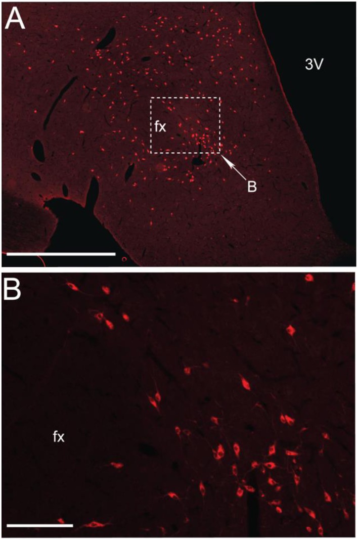
Figure 1.
MCHergic neurons are located in the hypothalamus. (A) Low magnification photomicrographs that exhibit MCHergic neurons at the tuberal level of the hypothalamus of the cat. (B) The inset in (A) is shown at higher magnification. This photomicrograph shows MCHergic neurons of the perifornical region. The photomicrographs were taken from 20 μm -thick sections that were processed for immunofluorescence. Fx, fornix; 3 V, third ventricle. Calibration bars: (A) 1 mm; (B) 100 μm. Original microphotographs taken from the data set of Torterolo et al. (2006).

